PROCEDURES PAGE
-
IT PROCEDURE FOR INSERTING BACTERIA
The IT was done by weighing each mouse and giving an appropriate amount
of Pentobarbital to sedate the mouse. The trachea was exposed and
the bacteria inserted. The trachea and the hole were clipped shut.
-
PENTOBARBITAL OVERDOSE PROCEDURE
The mice were picked up and 2 ml of Pentobarbital was injected in the mouse.
The injection was timed so that the heart would still be pumping when the
rest of the procedure is followed. 16 mice at each time were sacrificed
at each time point in this manner.
-
BRONCHIO-ALVEOLAR LAVAGE (BAL) PROCEDURE
The lungs of the mice in this experiment were lavaged to remove any cells
that were present inside the lung fluid. In lung infections, several
types of leukocytes enter the lung. The cells we are interested in
this experiment are the neutrophils and macrophages. The BAL procedure
follows:
1. The trachea was cleared by removing the clip.
2. A hole was cut in the trachea and a cathode inserted.
3. A knot was tied on the cathode so fluid would not leak.
4. .8 ml PBS with .5% EDTA was inserted and removed from the lungs
twice (1.6ml total). The EDTA serves as a detergent so cells don't
clot in the PBS solution.
5. The cathode is removed.
-
STEPS AFTER BAL INCLUDING CYTO SPINS
NOTE: The 4 and 24 hour
time plots were not disturbed to perform these procedures. Fluid
was flash frozen until all mice of that particular time plot were ready.
1. The fluid obtained from the BAL was centrifuged to concentrate cells.
(8 min at 1600)
2. The supernatant was removed. The cells were resuspended in
400uL PBS.
3. The cells were counted and the number in each tube readjusted to
1X10EE6 cells/ml.
4. 100 uL of each suspension was used to make cyto spin preparations.
Duplicates prepared.
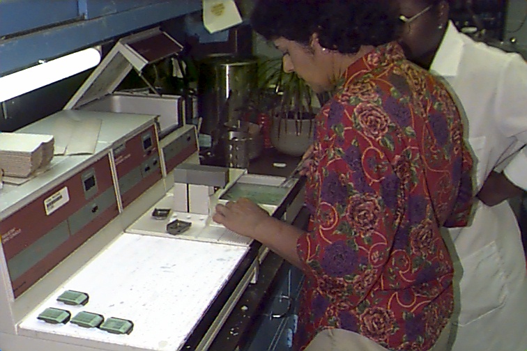
CYTO SPIN PREPARATION
1. Insert slide, filter, funnel into metal holder.
2. Pour suspension from step 4 above into funnel.
3. Spin at 500 rpm for 2 min.
-
RETRO-ORBITAL
BLEED AND BLOOD SMEAR PROCEDURES
1. After sacrificing mouse, a pasteur pipette was used to induce bleeding
from behind the eye of the mouse.
2. One drop of this blood was placed on a slide and smeared down the
slide using another slide.
3. This will show the cells that were present in the peripheral blood
of both types of mice.
-
LUNG PROFUSION AND REMOVAL
1. The heart was exposed.
2. A dark spot on the heart tissue signifies the right ventricle.
3 ml of PBS was injected here.
3. A successful profusion will show the lungs becoming white.
At this point, the lungs were removed and snap frozen (-80) for MPO and
CFU assays.
1. Dip each slide four times in the fixative.
2. Dry slide on paper towel and dip in eotoxin (pink) four times.
3. Dry slide again and dip in hematoxilin (purple) two times.
4. Examine slide under microscope for appropriate staining. Adjust
procedure accordingly.
-
PARAFIN EMBEDDING PROCEDURE
1. The samples were loaded into small plastic holders.
2. The lungs were flooded with gradual increases in alcohol concentration.
3. Metal holders were obtained and partially filled with parafin.
4. The samples were then taken out of the plastic holders and placed
in the metal ones avoiding the sides of the holder.
5. A plastic cover was put on and parafin was allowed to fill the holder.
6. These were allowed to cool at -3 degrees Celsius.
7. Once cool, the metal holder was removed.

-
MICROTOME CUTTING AND
STAINING PROCEDURE
1. A microtome was used to cut 3 .3 uM sections from the center of the
parafin block.
2. This was put on a slide and exposed to decreasing concentrations
of ethanol until dH2O.
3. Hema staining procedures were performed.
-
COLONY FORMING ASSAY (CFU) PROCEDURE
1. This procedure was performed with all 8 samples of lung mince.
1:10 serial dillutions were made in a 96 well plate. Each well of
the plate was filled with 90 uL PBS. 10 uL of sample mince solution was
placed in in the first row of the plate.
2. 10 uL of this was removed and put into the second row. 10
uL was removed from the second and put into the third row. This was
repeated for all 8 rows.
3. 10 uL from each dillution was plated on blood agar plates.
4. Each bacteria will form an independent colony. Counting the
number of colonies will tell how many bacterial cells were present in the
initial lung mince. Serial dillutions are made so that bacteria are
able to be counted if there are too many in one dillution.
-
MYLO-PEROXIDASE ASSAY (MPO) PROCEDURE
1. 100 uL of PBS slurry was put into 1.5 ml eppendorf.
2. An equal volume of 2X MPO homogenization buffer was adder to the
slurry. Warm buffer to 37C before addition.
3. Solution was sonicated three times for ten seconds each.
4. The solution was spinned for 15 min, 4C, at top speed.
5. Store at 4C.
6. ELISA READER SETUP: 490 nm, 2 min run time, shake=low, time=5 sec,
read int.=40 sec.
7. 10 uL of supernatant was put into the bottom of a 96 well plate.
8. The plate was put on the ELISA reader.
9. 140 uL of assay buffer was adder to the plate using multi-channel
pippette. The reading was started immediately.
10. SOLUTIONS NEEDED IN MPO:
1x Homogenization buffer
.25ug HTAB
50 ul .5 M EDTA
22 ml 1 M monobasic potassium phosphate
3.1 ml 1 M dibasic potassium phosphate
dH20 to Q.S. to 50 ml.
MPO Assay buffer
2.2 ml 1M monobasic potassium phosphate
310ul 1 M dibasic potassium phosphate
41.7 ul H2O2 (dilute 30% stock with 1:100 sterile
H2O)
420ul ODH (10mg/ml)
Q.S. to 25 ml with dH2O

